CANCER AWARENESS
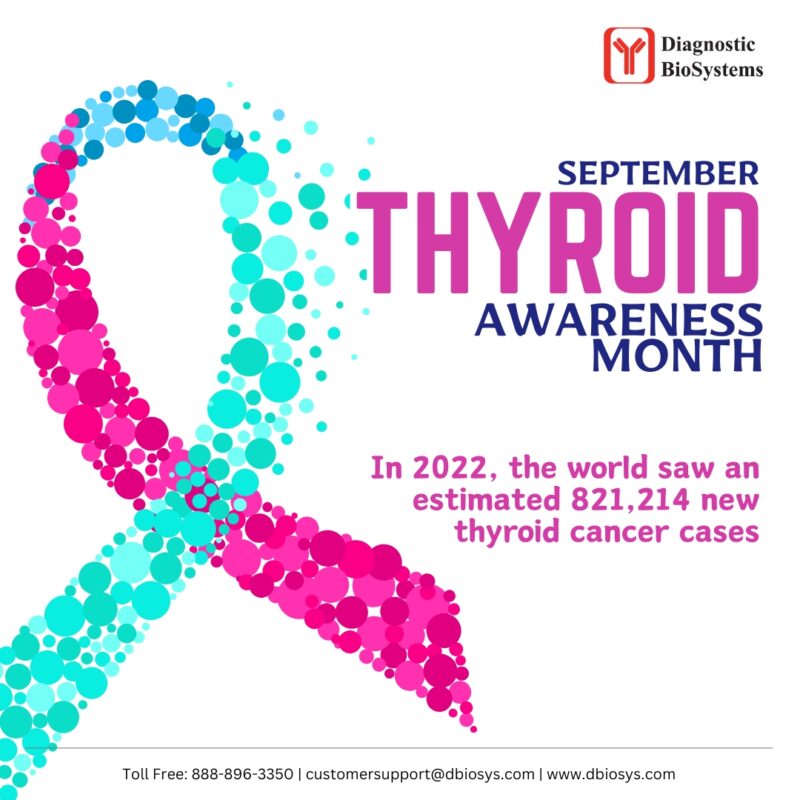
Thyroid Cancer Awareness
Thyroid Cancer Awareness
Thyroid nodules may be detected on physical examination or diagnosed incidentally on diagnostic imaging studies. The prevalence of palpable thyroid nodules has been reported to be approximately 5% in women and 1% in men who live in iodine-sufficient areas of the world. Current guidelines suggest that between 7%and 15%of thyroid nodules are malignant. Nodules 1 cm or larger should only be fine needle aspiration biopsied when ultrasound characteristics are concerning for malignant disease.
A comprehensive genomic analysis of 50734 indeterminate nodules found that 65.3% tested negative for malignancy, 33.9%tested positive, 0.6%were positive for parathyroid tissue, and 0.2%were positive for medullary thyroid cancer. Fine needle aspiration biopsies show no morphologic patterns of typical histology of thyroid cancers. Immunohistopathology (IHC) usually plays a very important role in making a diagnosis for thyroid cancers.
The immunohistochemical panels for thyroid fine needle aspiration biopsies are as follows:
- Follicular cell-derived lesions: PAX 8 (+, best), TTF-1(+), Thyroglobulin (+, but difficult to interpret)
- Papillary Thyroid carcinoma: CK 19 (+), MIB-1 (+, Nuclear), Beta-Catenin (+, cell membrane), ER(-), PR(-)
- Hyalinizing trabecular tumor: MIB-1 (+, membrane), Type IV collagen (+, hyaline material)
- Cribriform morular thyroid carcinoma: Beta-Catenin (+, nuclear and cytoplasm), ER(+), PR (+)
- Medullary thyroid carcinoma: Calcitonin (+), CEA (+), Chromogranin A (+), NSE (+), synaptophysin (+)
- Intrathyroid thymic carcinoma: CD5 (+), p16 (+), p40 (+), HMW CK (+), CD117 (+)
- Parathyroid adenoma: GATA3 (+), parathyroid hormone (+), Chromogranin A (+)
Metastatic carcinoma
- Renal cell carcinoma: PAX8 (+), CD10 (+)
- Lung carcinoma: Napsin A (+), TTF-1 (+)
- Breast carcinoma: GATA3 (+), ER(+), PR (+), Her2 (+).


