CD43 T-Cell
Description
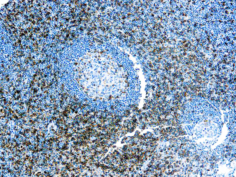
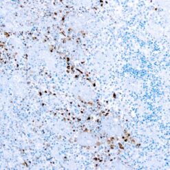
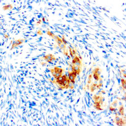
It recognizes a cell surface glycoprotein of 95/115/135kDa (depending upon the extent of glycosylation), identified as CD43 [Workshop IV]. Epitope of MAb Bra7G is clearly different from that of MAb DF-T1, called b as opposed to a for DF-T1. 70-90% of T-cell lymphomas and from 22-37% of B-cell lymphomas express CD43. No reactivity has been observed with reactive Bcells. So a B-lineage population that co-expresses CD43 is highly likely to be a malignant lymphoma, especially a low-grade lymphoma, rather than a reactive B-cell population. When CD43 antibody is used in combination with anti-CD20, effective immunophenotyping of the lymphomas in formalinfixed tissues can be obtained. Co-staining of a lymphoid infiltrate with antiCD20 and anti-CD43 argues against a reactive process and favors a diagnosis of lymphoma.
Additional information
| Clone | DF-T1 |
|---|---|
| Isotype | IgG1, kappa |
| Immunogen | BALB/C mice were immunized with myeloblast cell line KG1. |
| Species | Mouse |
| Cellular Localization | cell membrane |
| Positive Control Tissue | Tonsil |
| Pretreatment | EDTA Buffer pH 8.0 (Manual/ Montage) |
| Incubation & Temperature | 30 min @ RT |
| Intended Use | IVD |
| Detection System | PolyVue™ Plus – Two Step Detection System or Montage PolyVue Plus™ Auto Detection System for Montage 360 System or HighLighter core kit for HighLighter Staining System |
| Description/Type | Conc Mouse Monoclonal Antibody |
| Format | This product is supplied as a purified immunoglobulin and contains sodium azide as a preservative. |
DATASHEETS & SDS
DATASHEETS & SDS
| Download Datasheet |
| Download SDS Sheet – OSHA |
REFERENCES
REFERENCES
- Stross WP, et. al. Journal of Clinical Pathology, 1989, 42(9):953-61.
Reviews (0)
Only logged in customers who have purchased this product may leave a review.





Reviews
There are no reviews yet.