MyoD1 (5.2F)
Description
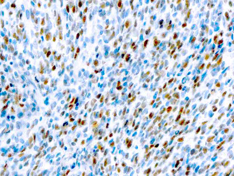
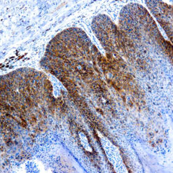
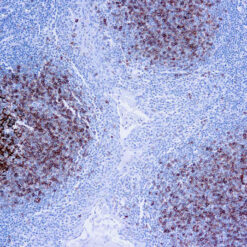
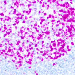
Recognizes a phosphor-protein of 45kDa, identified as MyoD1. The epitope of this MAb maps between amino acid 180-189 in the C-terminal of mouse MyoD1 protein. It does not cross react with myogenin, Myf5, or Myf6. Antibody to MyoD1 labels the nuclei of myoblasts in developing muscle tissues. MyoD1 is not detected in normal adult tissue, but is highly expressed in the tumor cell nuclei of rhabdomyosarcomas. Occasionally nuclear expression of MyoD1 is seen in ectomesenchymoma and a subset of Wilm’s tumors. Weak cytoplasmic staining is observed in several non-muscle tissues, including glandular epithelium and also in rhabdomyosarcomas, neuroblastomas, Ewing’s sarcomas and alveolar soft part sarcomas.
Additional information
| Catalog No. | Mob278 Concentrated, PDM120 Prediluted |
|---|---|
| Clone | 5.2F |
| Isotype | IgG2a |
| Immunogen | Recombinant mouse MyoD1 protein. |
| Species | Mouse |
| Cellular Localization | Nuclear |
| Positive Control Tissue | Rhabdomyosarcoma |
| Pretreatment | EDTA Buffer pH 8.0 |
| Incubation & Temperature | 30 min @ RT |
| Intended Use | IVD |
| Detection System | PolyVue Plus – Two Step Detection System or Montage PolyVue Plus Auto Detection System for Montage 360 System |
| Description/Type | Mouse Monoclonal Antibody |
| Format | Purified immunoglobulin |
DATASHEETS & SDS
DATASHEETS & SDS
| Download Datasheet |
| Download SDS Sheet – OSHA |
REFERENCES
REFERENCES
- Thulasi R et. al. Cell Growth and Differentiation, 1996, 7(4):531-41.
- Wesche WA et. al. American Journal of Surgical Pathology, 1995,
19(3):261-9. - Parham DM et. al. Acta Neuropathologica, 1994, 87:605-11
Reviews (0)
Only logged in customers who have purchased this product may leave a review.






Reviews
There are no reviews yet.