p53 (DO-7)
Description
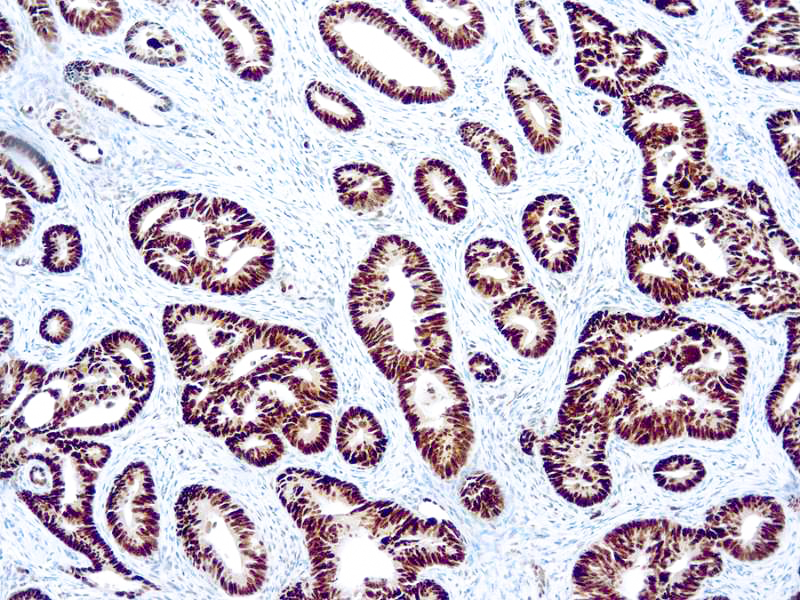
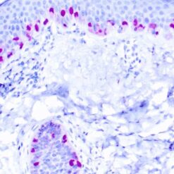
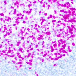
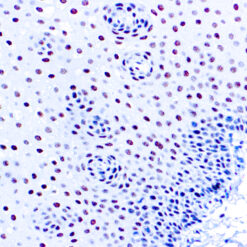
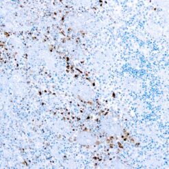
This antibody reacts with the mutant as well as the wild form of p53. Its epitope maps within N-terminus (aa 37-45) of p53. p53 is a tumor suppressor gene expressed in a wide variety of tissue types and is involved in cell growth, replication, and apoptosis. It binds to MDM2, SV40 T antigen and human papilloma virus E6 protein. Positive nuclear staining with p53 antibody has been reported to be a negative prognostic factor in breast carcinoma, lung carcinoma, colorectal, and urothelial carcinoma. Anti-53 positivity has also been used to differentiate uterine serous carcinoma from endometrioid carcinoma as well as to recognize intratubular germ cell neoplasia. Mutations involving p53 are found in a wide variety of malignant tumors, including breast, ovarian, bladder, colon, lung, and melanoma.
Additional information
| Catalog No. | Mob082 Concentrated, PDM013 Prediluted |
|---|---|
| Clone | DO7 |
| Isotype | IgG2b, kappa |
| Immunogen | Recombinant human wild type P53 protein expressed in E. coli. |
| Species | Mouse |
| Cellular Localization | Nuclear |
| Positive Control Tissue | Colon carcinoma |
| Pretreatment | Citrate Buffer pH 6.0 |
| Incubation & Temperature | 30 min @ RT |
| Intended Use | IVD |
| Detection System | PolyVue™ Plus – Two Step Detection System or Montage PolyVue Plus™ Auto Detection System for Montage 360 System or HighLighter core kit for HighLighter Staining System |
| Description/Type | Mouse Monoclonal Antibody |
| Format | This product is supplied as a cell culture supernatant and contains sodium azide as a preservative |
DATASHEETS & SDS
DATASHEETS & SDS
| Download Datasheet |
| Download SDS Sheet – OSHA |
REFERENCES
REFERENCES
- Vojtesek B et al. 1992. J. Immunol. Methods. 151(1-2): 237-44.
- Stephen CW et al. 1995. J Mol. Biol. 248(1): 58-78.
Reviews (0)
Only logged in customers who have purchased this product may leave a review.





Reviews
There are no reviews yet.