DIAGNOSTIC BIOSYSTEMS INC.
TESTIMONIALS

Altium International SRL is the Authorized Distributor of Agilent Technologies in Romania with its headquarter located in Bucharest. We are providing high quality solutions to customers at universities, research institutes, clinical and anatomic pathology laboratories for more than 20 years.
Diagnostic BioSystems is our reliable business partner for more than two years providing us with high quality antibodies that complement our portfolio of Agilent Technologies (DAKO IHC products) for our Anatomic Pathology Division.
Altium International SRL’ customers are very much satisfied with the quality and performance of Diagnostic Biosystems’ primary antibodies. Our business with Diagnostic BioSystems has been growing steadily over the last two years. DBS antibodies are easy to optimize on both, our DAKO Omnis as well as on DAKO Autostainers.
We have a broad customer base at public and private hospitals and institutions who integrated DBS antibodies in their daily routine work after a successful validation on our instruments.
Meanwhile we carry more than 50 different antibodies from DBS in our portfolio that is constantly increasing. We appreciate the consistent high quality, the short-term availability and long shelf life of the products from Diagnostic BioSystems as well as the commitment to certify all of its antibodies as IVDR.
Our collaboration is excellent; we are getting instant technical and logistic support whenever required. We highly recommend Diagnostic BioSystems as a business partner in the field of Immunohistochemistry.
Sincerely,


YouTube Link: https://youtu.be/yFYSwmD6lgE
I have been using DBS antibodies for some time now and I must say that the quality is really very good.
Their products are all FDA approved and I can assure you that the quality of the antibodies are very good.
I have used their common markers and it works quite well, and the staining is very fine and crisp.
DBS antibodies also work well in other automation too and that’s an added advantage for the automated users. Their customer service and support team are always on their toes to help and support whenever required.
I would like to thank Diagnostic BioSystems for providing such quality IHC products on time everytime.
Review of Mouse/Rabbit PolyVue Plus™ HRP/DAB Detection System (PVP250D)
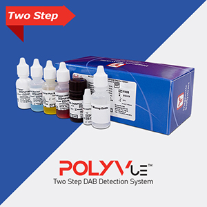
Performance of Mouse/Rabbit PolyVue Plus™ HRP/DAB Detection System in the immunohistochemical analysis of Cytokeratin 5 (XM26) and p16 (JC2) on melanoma tissue.
Materials and Methods:
Reagents obtained from Diagnostic BioSystems:
Cytokeratin 5 (XM26) and p16 (JC2) concentrated format. Mouse/Rabbit PolyVue Plus™ HRP/DAB Detection System, Montage™ Hematoxylin.
Staining was performed manually as well as on the autostainer intelliPATH FLX®(Biocare Medical).
Both methods lead to comparable results.
Pre-Treatment Module PT Link (Agilent/ Dako) was used for heat-induced epitope retrieval.
Protocol:
Tissue sections were de-waxed in an oven for 1 hour at 65ºC. For antigen retrieval, sections were incubated in the Dako PT Link, using Citrate buffer pH 6.0 for CK5 (XM26) and EDTA buffer pH 8.0 for p16 (JC2) for 15 min at 95ºC. After cooling down to room temperature, sections were washed with 3 times with Immuno Wash Buffer. Then, the staining process was applied according to the staining protocols specified by the Instructions for Use of the supplier. Both antibodies were diluted a at a ratio of 1:50 and incubated for 30 min at room temperature. The sections were developed with DAB/Plus™ for 5 min at room temperature. As a nuclear counter stain, Montage™ Hematoxylin was incubated for 5 min at room temperature, followed by dehydration and coverslip mounting.
Results:
The results we achieved with the Mouse/Rabbit PolyVue Plus™ HRP/DAB Detection System are optimal for both antibodies:
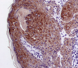 IHC image of CK5 (XM26) staining in melanoma formalin fixed paraffin embedded tissue section.The tumor cells are stained strongly in cytoplasm and lymphocytes are completely not stained as an internal negative control.
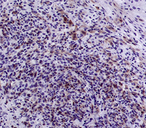
IHC image of p16 (JC2) staining in melanoma formalin fixed paraffin embedded tissue section. p16 stained nuclei and cytoplasm of melanoma cells and the background lymphocytes are not positive at all as an internal negative control.
Conclusion:
The described method reduced background artefacts significantly showing expressed tissue sections in a clean and clear manner. The PolyVue Plus Detection System is well suited for both mouse and rabbit antibodies, so it is an excellent kit for efficient and fast staining. It is a preferred alternative for routine laboratory work since it is quick and easy to use.
Customer Name
GENNOVA SCIENTIFIC S.L.
c/ Johann Gutenberg, 4F.
P.I. El Cáñamo I. 41300
San José de la Rinconada.
Sevilla, Spain

Diagnostic Biosystems exhibits a meticulous focus on quality and meaningful validation for their antibody products. Since 2010, when ours’ and DBS’ relationship began, our clients have consistently expressed delight with the reliability, technical expertise and custom service that support their products. The company portfolio and the scientific team behind it have built an exceptional organization truly in service to scientists doing cutting-edge biological research in labs around the world.
John Mountzouris, Ph.D.
Chief Scientific Officer
Abcepta

We trust in Diagnostic Biosystems’ Highlighter for the study of Immunohistochemistry HRP/DAB, valuing their personalized technical support and permanent commitment to accompanying us in our growth, both of which are vital for the welfare of our patients. We appreciate the excellent work of their entire team of expert professionals and the quality technology offered, which guarantees optimal results.
EXAMEDIC
Laboratorio de Anatomía Patológica
Dra. Carolina Henestrosa J.
Dra. Lily Márquez S.


