


 1")
Description
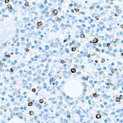
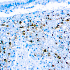
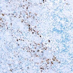
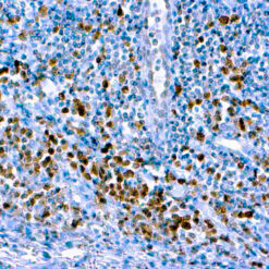
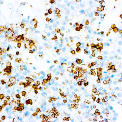
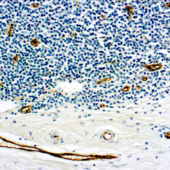
This antibody reacts with a glycoprotein of 110 kDa, expressed as intracytoplasmic molecule. It stains macrophages in a wide variety of tissues. Myeloid precursor cells and peripheral granulocytes are negative. The PGM1 differs from EMB11 (CD68) because of its non-reactivity with granulocytes and their precursor cells.
Additional information
| Clone | PG-M1 |
|---|---|
| Isotype | IgG3, kappa |
| Immunogen | BALB/C mice were immunized with Gauchers cells. |
| Species | Mouse |
| Cellular Localization | cell membrane |
| Positive Control Tissue | Tonsil |
| Pretreatment | HistoZyme (Manual/ Montage) |
| Incubation & Temperature | 30 min @ RT |
| Intended Use | IVD |
| Detection System | PolyVue™ Plus – Two Step Detection System or Montage PolyVue Plus™ Auto Detection System for Montage 360 System or HighLighter core kit for HighLighter Staining System |
| Description/Type | Mouse Monoclonal Antibody |
| Format | This product is supplied as a tissue culture supernatant and contains sodium azide as a preservative. |
DATASHEETS & SDS
DATASHEETS & SDS
| Download Datasheet |
| Download SDS Sheet – OSHA |
REFERENCES
REFERENCES
- Horny et al. Human Pathol 24; 35S, 1993.
- Kaiserling et al. Modern Path& 6: 463, 1993.
- Thiele et al. Virchows Archly A. Pathol Mat 421: 33, 1992.
Reviews (0)
Only logged in customers who have purchased this product may leave a review.


Reviews
There are no reviews yet.