p63/p504s Cocktail
Description
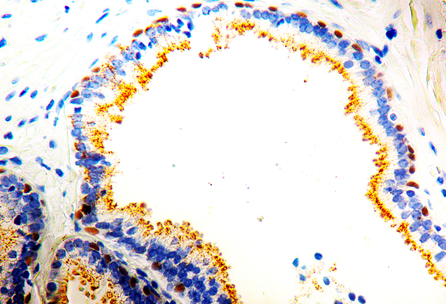
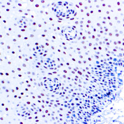
p63 is a homolog of the tumor suppressor p53. It is identified in basal cells in the epithelial layers of a variety of tissues, including epidermis, cervix, urothelium, breast and prostate. p63, vital for the development of the prostate and selectively expressed by normal prostate basal cells, has been shown useful in the differential diagnosis of benign prostatic lesions and prostatic carcinoma. p63 has also been shown to be highly sensitive and specific for detecting lung squamous cell carcinomas of the breast. p63 staining can be identified in myoepithelial cells of normal ducts. The expression of p504S protein is found in prostatic adenocarcinoma. It stains premalignant lesions of prostate: high-grade prostatic intraepithelial neoplasia (PIN) and atypical adenomatous hyperplasia. P504S can be used as a positive marker for PIN.
Additional information
| Catalog No. | PDRMP001 |
|---|---|
| Clone | DBR 16.1 / Polyclonal |
| Species | Rabbit |
| Isotype | IgG / N/A |
| Immunogen | Recombinant human p63 protein fragment + Synthetic human AMACR peptide |
| Cellular Localization | Nuclear and Cytoplasmic |
| Positive Control Tissue | Prostate |
| Pretreatment | EDTA Buffer pH 8.0 |
| Incubation & Temperature | 30 min @ RT |
| Intended Use | IVD (Outside Europe) |
| Detection System | PolyVue™ Plus – Two Step Detection System or Montage PolyVue Plus™ Auto Detection System for Montage 360 System |
| Description/Type | Rabbit Mono/Polyclonal Antibody |
| Format | This product is supplied as a purified immunoglobulin and contains sodium azide as a preservative. |
DATASHEETS & SDS
DATASHEETS & SDS
| Download Datasheet |
| Download SDS Sheet – OSHA |
REFERENCES
REFERENCES
I. Yang A, et al. p63, a p53 homolog at 3q27–29, encodes multiple products with transactivating, death-inducing, and dominant-negative activities. Mol Cell. 1998 Sep; 2(3):305-16.
II. Mukhopadhyay S, et al. Subclassification of non-small cell lung carcinomas lacking morphologic differentiation on biopsy specimens: Utility of an immunohistochemical panel containing TTF-1, napsin A, p63, and CK5/6. Am J Surg Pathol. 2011 Jan; 35(1):15-25.
III. Tacha D, et al. A six antibody panel for the classification of lung adenocarcinoma versus squamous cell carcinoma. Appl Immunohistochem Mol Morphol. 2012 May; 20 (3):201-7.
IV. Terry J, et al. Optimal immunohistochemical markers for distinguishing lung adenocarcinomas from squamous cell carcinomas in small tumor samples. Am J Surg Pathol. 2010 Dec; 34(12):1805-11.
V. Pu RT, et al. Utility of WT-1, p63, MOC31, mesothelin, and cytokeratin (K903 and CK5/6) immunostains in differentiating adenocarcinoma, squamous cell carcinoma, and malignant mesothelioma in effusions. Diagn Cytopathol. 2008 Jan; 36(1):20-5.
VI. Lerwill MF. Current practical applications of diagnostic immunohistochemistry in breast pathology. Am J Surg Pathol. 2004 Aug; 28(8):1076-91
Reviews (0)
Only logged in customers who have purchased this product may leave a review.






Reviews
There are no reviews yet.