
Immunohistochemistry (IHC) is a powerful technique that uses the specific binding of an antibody to an antigen to detect and localise specific antigens in cells and tissues, which is typically detected and examined using a light microscope. With the advent of antigen retrieval methods that allow it to be performed conveniently on tissue sections during manual staining along with automated methods for high volume processing with reproducibility, IHC has become an essential ancillary technique in clinical diagnostics in anatomic pathology.
Diagnostic BioSystems is a leading developer and provider of speciality Immunohistochemistry reagents. DBS offers a wide range of products including Rabbit polyclonals and monoclonals and Mouse monoclonal antibodies, detection systems, instrumentation, and ancillary reagents with global quality.
Get in touch
Please fill out the below form to get in touch with us. We’ll be sure to get back to you as soon as we can.
PRODUCTS BY CATEGORY
 1")
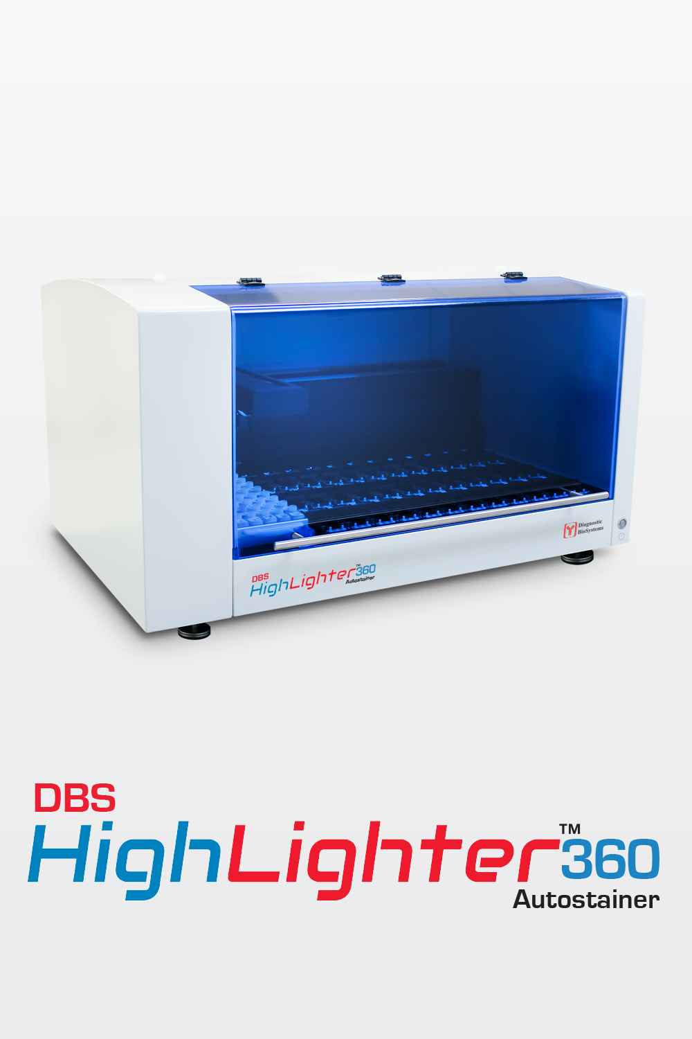
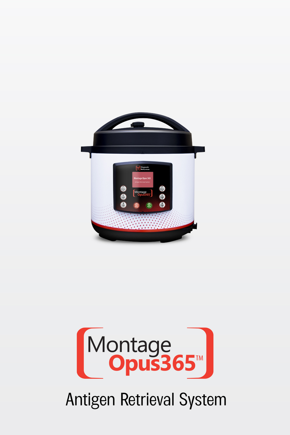
Immunohistochemistry (IHC) PRODUCTS
 7")
 8")
 9")
 10")
 11")
 12")
 13")
 14")
 15")
 16")
 17")
 18")
 19")
 20")
 21")
 22")
 23")
 24")
 25")
 26")
 27")
 28")
 29")
 30")
 31")
 32")
 33")
 34")
 35")
 36")
 37")
 38")
 39")
 40")
 41")
 42")
 43")
 44")
 45")
 46")
 47")
 48")
 49")
 50")
 51")
 52")
 53")
 54")
 55")
 56")
 57")
 58")
 59")
 60")
 61")
 62")
 63")
 64")
 65")
 66")
 67")
 68")
 69")
 70")
 71")
 72")
 73")
 74")
 75")
 76")
 77")
 78")
 79")
 80")
 81")
 82")
 83")
 84")
 85")
 86")
 87")
 88")
 89")
 90")
 91")
 92")
 93")
 94")
 95")
 96")
 97")
 98")
 99")
 100")
 101")
 102")
 103")
 104")
 105")
 106")
 107")
 108")
 109")
 110")
 111")
 112")
 113")
 114")
 115")
 116")
 117")
 118")
 119")
 120")
 121")
 122")
 123")
 124")
 125")
 126")
 127")
 128")
 129")
 130")
 131")
 132")
 133")
 134")
 135")
 136")
 137")
 138")
 139")
 140")
 141")
 142")
 143")
 144")
 145")
 146")
 147")
 148")
 149")
 150")
 151")
 152")
 153")
 154")
 155")
 156")
 157")
 158")
 159")
 161")
 162")
 163")
 164")
 165")
 166")
 167")
 168")
 169")
 170")
 171")
 172")
 173")
 174")
 175")
 176")
 177")
 178")
 179")
 180")
 181")
 182")
 183")
 184")
 185")
 186")
 187")
 188")
 189")
 190")
 191")
 192")
 193")
 194")
 195")
 196")
 197")
 198")
 199")
 200")
 201")
 202")
 203")
 204")
 205")
 206")
 207")
 208")
 209")
 210")
 211")
 212")
 213")
 214")
 215")
 216")
 217")
 218")
 219")
 220")
 221")
 222")
 223")
 224")
 226")
 227")
 228")
 230")
 231")
 232")
 233")
 234")
 235")
 236")
 237")
 238")
 239")
 240")
 241")
 242")
 243")
 244")
 245")
 246")
 247")
 248")
 249")
 250")
 251")
 252")
 253")
 254")
 255")
 256")
 257")
 259")
 260")
 261")
 262")
 263")
 264")
 265")
 266")
 267")
 268")
 269")
 270")
 271")
 272")
 273")
 274")
 275")
 276")
 277")
 278")
 279")
 280")
 281")
 282")
 283")
 284")
 285")
 286")
 287")
 288")
 289")
 290")
 291")
 292")
 293")
 294")
 295")
 296")
 297")
 298")
 299")
 300")
 301")
 302")
 303")
 304")
 305")
 306")
 307")
 308")
 309")
 310")
 311")
 312")
 313")
 314")
 315")
 316")
 317")
 318")
 319")
 320")
 321")
 323")
 324")
 325")
 327")
 329")
 330")
 331")
 332")
 333")
 334")
 335")
 336")
 337")
 338")
 339")
 340")
 341")
 342")
 343")
 344")
 345")
 346")
 347")
 348")
 349")
 350")
 351")
 352")
 353")
 354")
 355")
 356")
 357")
 359")
 360")
 361")
 362")
 363")
 364")
 365")
 366")
 367")
 368")
 369")
 370")
 371")
 372")
 373")
 374")
 375")
 376")
 377")
 378")
 379")
 380")
 381")
 382")
 383")
 384")
 385")
 386")
 387")
 388")
 389")
 390")
 392")
 393")
 394")
 395")
 396")
 397")
 398")
 399")
 400")
 401")
 402")
 403")
 404")
 405")
 406")
 407")
 408")
 409")
 410")
 412")
 413")
 414")
 415")
 416")
 417")
 418")
 421")
 422")
 423")
 424")
 425")
 426")
 427")
 428")
 429")
 430")
 431")
Overview and Definition of immunohistochemistry
Immunohistochemistry (IHC) is the most common application of immunostaining. It is the process of identifying antigens (proteins) in cells of a tissue section by utilising the principle of antibodies binding specifically to antigens in biological tissues. IHC derives its name from the words “immuno,” which refers to the antibodies used in the procedure, and “histo,” which means “tissue” (compare to immunocytochemistry). Albert Coons conceptualised and first implemented the procedure in 1941.
Visualizing an antibody-antigen interaction can be done in a variety of ways, the most common of which are:
- Chromogenic immunohistochemistry (CIH), in which an antibody is conjugated to an enzyme, such as peroxidase (the combination is known as immunoperoxidase), which can catalyse a color-producing reaction.
- Immunofluorescence, in which an antibody is linked to a fluorophore, such as fluorescein or rhodamine.
People also ask - Immunohistochemistry (IHC) frequently asked questions (FAQs)
What is the process of immunohistochemistry?
Immunohistochemistry (IHC) is the most common application of immunostaining. It is the process of identifying antigens (proteins) in cells of a tissue section by utilising the principle of antibodies binding specifically to antigens in biological tissues. IHC derives its name from the words “immuno,” which refers to the antibodies used in the procedure, and “histo,” which means “tissue” (compare to immunocytochemistry).
What diseases can be diagnosed by immunohistochemistry?
IHC is widely used for diagnosis of cancers; specific tumor antigens are expressed de novo or up-regulated in certain cancers.
What is immunohistochemistry positive?
immunohistochemistry test (IHC Test) is used to diagnose cancer by detecting specific kinds of antibodies attached to antigens in a cell. This test helps to identify tumors; also, it can distinguish whether or not the tumor is benign or malignant.
If the test is negative, it means that the laboratory did not find any changes in the proteins of the tumor.
If the test is positive, the result means that the laboratory found a particular change in the proteins of the tumor
What are the methods of immunohistochemistry?
Immunohistochemistry use different staining procedures such as one step direct method, ABC methods, two-step indirect method and Tyramide signal amplification.
immunohistochemistry vs immunofluorescence
The three staining techniques differ in the sample/tissue type:
- immunofluorescence is commonly used to stain microbiological cells
- immunohistochemistry is commonly used to stain sections of biological tissue
- immunocytochemistry is commonly used to stain intact cells removed from extracellular matrix